
Search
- Page Path
- HOME > Search
Original Articles
- Clinical Study
- Relationships between Thigh and Waist Circumference, Hemoglobin Glycation Index, and Carotid Plaque in Patients with Type 2 Diabetes
- Myung Ki Yoon, Jun Goo Kang, Seong Jin Lee, Sung-Hee Ihm, Kap Bum Huh, Chul Sik Kim
- Endocrinol Metab. 2020;35(2):319-328. Published online June 24, 2020
- DOI: https://doi.org/10.3803/EnM.2020.35.2.319
- 8,368 View
- 145 Download
- 4 Web of Science
- 3 Crossref
-
 Abstract
 PDF
PDF PubReader
  ePub
ePub - Background
This study investigated the relationships of thigh and waist circumference with the hemoglobin glycation index (HGI) and carotid atherosclerosis in patients with type 2 diabetes.
Methods
This observational study included 3,075 Korean patients with type 2 diabetes, in whom anthropometric measurements and carotid ultrasonography were conducted. HGI was defined as the measured hemoglobin A1c (HbA1c) level minus the predicted HbA1c level, which was calculated using the linear relationship between HbA1c and fasting plasma glucose levels. Carotid atherosclerosis was defined as a clearly isolated focal plaque or focal wall thickening >50% of the surrounding intima-media thickness.
Results
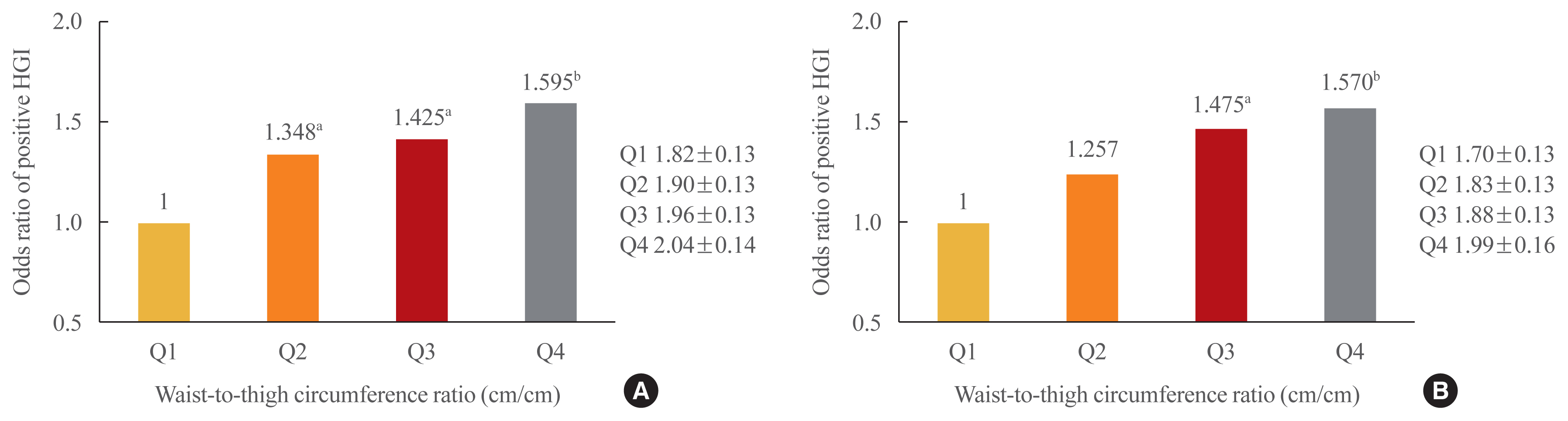
The frequency of a positive HGI decreased with increasing thigh circumference in men and increased with increasing waist circumference in women after adjusting for potential confounding variables. Thigh and waist circumference had a combined augmentative effect on the likelihood of positive HGI, which was dramatically higher in patients in higher waist-to-thigh ratio quartiles (adjusted odds ratios for the highest compared to the lowest quartile: 1.595 in men and 1.570 in women). Additionally, the larger the thigh circumference, the lower the risk of carotid atherosclerosis, although in women, this relationship lacked significance after adjustment for potential confounders.
Conclusion
HGI was associated with thigh circumference in men and waist circumference in women. In addition, the combination of low thigh circumference and high waist circumference was strongly associated with a higher HGI in Korean patients with type 2 diabetes. In particular, thigh circumference was associated with carotid atherosclerosis in men. However, further longitudinal studies are warranted. -
Citations
Citations to this article as recorded by
- Association between hemoglobin glycation index and subclinical myocardial injury in the general population free from cardiovascular disease
Zhenwei Wang, Yihai Liu, Jing Xie, Nai-Feng Liu
Nutrition, Metabolism and Cardiovascular Diseases.2022; 32(2): 469. CrossRef - Association of Hemoglobin Glycation Index With Contrast-Induced Acute Kidney Injury in Patients Undergoing Coronary Angiography: A Retrospective Study
Zhezhe Chen, Duanbin Li, Maoning Lin, Hangpan Jiang, Tian Xu, Yu Shan, Guosheng Fu, Min Wang, Wenbin Zhang
Frontiers in Physiology.2022;[Epub] CrossRef - Associations of continuous glucose monitoring-assessed glucose variability with intima-media thickness and ultrasonic tissue characteristics of the carotid arteries: a cross-sectional analysis in patients with type 2 diabetes
Naohiro Taya, Naoto Katakami, Tomoya Mita, Yosuke Okada, Satomi Wakasugi, Hidenori Yoshii, Toshihiko Shiraiwa, Akihito Otsuka, Yutaka Umayahara, Kayoko Ryomoto, Masahiro Hatazaki, Tetsuyuki Yasuda, Tsunehiko Yamamoto, Masahiko Gosho, Iichiro Shimomura, Hi
Cardiovascular Diabetology.2021;[Epub] CrossRef
- Association between hemoglobin glycation index and subclinical myocardial injury in the general population free from cardiovascular disease

- Estrogen Receptor Gene Polymorphism, Urinary Estrogen Metabolites and Bone Mineral Density in Korean Postmenopausal Women.
- Ji Hyun Lee, Sung Kil Lim, Young Jun Won, Seok Ho Kwon, Bong Soo Cha, Young Duk Song, Hyun Chul Lee, Kap Bum Huh
- J Korean Endocr Soc. 1996;11(4):468-478. Published online November 7, 2019
- 1,235 View
- 22 Download
-
Abstract
PDF
- Background
Estrogen status is important for maintaining the homeostasis of bone. Estrogen has direct effects on bone cells, through binding to the high-affinity estrogen receptor. Several recent studies suggest that there might be genetically determined variations in biosynthesis and function of estrogen receptor in postmenopausal osteoporosis. Also the main cause of postmenopausal osteoporosis is decreased level of serum estrogen, whereas there had been some suggestion that the remaining estrogen have some effect on bone metabolism after menopause. We investigated the relationship between estrogen receptor gene PvulI polymorphism and bone mineral density(BMD), and the relationship between 18 urinary metabolites of estrogen and BMD in Korean postmeno- pausal osteoporosis. Methods: We examined the PvuII polymorphism of the estrogen receptor gene in 5' upstream region and the first intron by restrietion frapnent length polymorphism analysis in 62 postmeno- pausal wornen, BMD was measured by DEXA. The urinary estrogen metabolites were determined by GC/MS(Gas Chromatography-Mass Spectrometry) at Korean Institute of Science and Techno- logy Doping Control Center. Results: BMD of the spine and the femoral neck correlated with body weight, height, body mass index as we expected. There was no polymorphism of PvuII restriction site on 5 upstream region of estrogen receptor gene. Whereas the prevalen~ee of the PP, Pp, pp genotype in the first intron of estrogen receptor was 12.9%, 45.2%, 41.9%, respectively. But, there was no correlation between PvuII genotype and the spinel and femoral neck BMD. 2(OH)E2 among 18 urinary metabolites of estrogen, showed a negative correlation with the spinal and femoral neck BMD(r =-0.2551, p<0.05, and r =-0.3341, p<0.01, respectively), and the ratio of 16a(OH)E2/2(OH)E1> revealed a positive correlation with the spinal BMD(r =0.3057, p<0.05). In stepwise multiple regression analysis, body weight, 2(OH)E2, 16a(OH)E1, 2(Meo)E1 were independent predictors of the spinal bone density, and body weight and 2(OH)E2 were independent predictors of the femoral neck bone density. Conclusion: These results suggested that restrietion fragment length polymorphism analysis of the estrogen receptor gene with PvuII restriction enzyme was not helpful for early detection of patients at risk of developing osteoporosis. However, the ratio of 16-hydroxylation to 2-hydroxylation of estrogen metabolism was reduced in postmenopausal women and high catecholestrogen formation might be a greater risk factor for osteoporosis.
Case Report
- A Case of Giant Adrenal Adenoma Presenting Primary Aldosteronism.
- Ji Hyun Lee, Bong Soo Cha, Moon Suk Nam, Young Duk Song, Sung Kil Lim, Hyun Chul Lee, Kap Bum Huh, Hyung Chan Suh, Young Hwa Choi, Jae Min Park, Jung Soo Park, Soon Won Hong, Dong Hwan Shin
- J Korean Endocr Soc. 1996;11(3):348-354. Published online November 7, 2019
- 1,295 View
- 25 Download
-
Abstract
PDF
- Primary aldosteronism is a syndrome chracterized by hypokalemic alkalosis and hypertension. Small sized adrenal cortical adenomas have been the major cause of this syndrome in most of the patients. However, if the adrenal mass is larger than 6cm in diameter and with irregular consistency, malignancy is more favored. We experienced a patient who had a giant adrenal adenoma with primary aldosteronism. A 24-year-old female presented with hypertension, hypokalemia, low plasma renin, and high plasrna aldosterone levels, was found to have a 6×5.5×5 cm sized left adrenal tumor by MRI. Her clinical laboratory feature did not revealed any evidence of Cushing's syndrome or pheochromocytoma. Preoperatively adrenal carcinoma presenting pure adrenal aldosteronism was suspected due to large size and heterogenous signal character of the adrenal mass in radiologic study. At operation well encapsulated, round giant adrenal tumor weighing 65gm(4.5×4×4 cm) was removed. There was no evidence of metastasis with return of adrenal function to normal after surgery. Benign adrenal adenoma was confirmed by the gross morphology and the histologic features.
Original Articles
- Clinical and Sellar MR Findings in Central Diabets Inspidus.
- Bong Soo Cha, Young Duk Song, Sung Kil Lim, Kyung Rae Kim, Hyun Chul Lee, Kap Bum Huh, Su Youn Nam, Eun Jig Lee, Sei Chang Oh, Byung Hee Lee, Dong Ik Kim
- J Korean Endocr Soc. 1996;11(3):285-292. Published online November 7, 2019
- 1,227 View
- 19 Download
-
Abstract
PDF
- Background
s: Diabetes insipidus(DI) is a clinical syndrome characterized by excretion of copious volumes of dilute urine combined with persistent intake of abnormally large quantities of fluid. Central DI, caused by lack of antidiuretic hormone(ADH), most often results from lesions in the hypothalamic-neurohypophyseal axis. Magnetic resonance(MR) imaging is particularly useful in documenting the presence of a structural lesion, as opposed to assigning a diagnosis of idiopathic DI for which only symptomatic therapy is prescribed. Recently, several reports have described a specific MR finding in central DI, that is absence of normal posterior pituitary bright spot(PPBS). Methods: We retrospectivesly studied the clinical and MR findings in 25 patients with central DI, diagnosed by warter deprivation test. Results: 1) The subjects included 17 males and 8 females, between the ages of 2 and 58 years. 2) 24-hour urine volumes were 2,340~13,750 mL, and mean urine osmolarity was 147.7±65.8 mOsm/kg. The 23 subjects diagnosed complete central DI by warter deprivation test. 3) We found that the most common cause of cntral DI was infiltrative lesions of hypothalmic-neurohypophyseal axis(60%). Germ cell tumor was the single leading cause in present study, accounting for 36% of cases. The other causes were found, including pituitary apoplexy, meningitis, and trauma. Idiopathic central DI accounted for 20% of all cases. 4) Growth hormone deficiency was the most common accompanying anterior pituitary deficit, and panhypopituitarism was found in 7 cases, Hyperprolactinernia was seen in 6 cases. 5) In all patients, PPBS on Tl weighted MR images were not observed. A thickened pituitary stalk was seen in 15 cases(9 patients with germ cell tumor, 3 patients with histiocytosis X, 1 patient with tuberculosis, 2 patients with unknown origin). Conclusion: In our results, the most common causes of central DI was suprasellar infiltrative lesions. MR is currently the imaging methods of choice in the evaulation of dysfunction of the hypothalamic-neurohypophyseal system in patients with central DI. A specific MR finding, that is loss of normal PPBS allows a confirmative diagnosis of central DI.
- Reduction of Central Dopamine Release in Hyperprolactinemia.
- Bong Soo Cha, Young Duk Song, Sung Kil Lim, Kyung Rae Kim, Hyun Chul Lee, Kap Bum Huh, Su Youn Nam, Eun Jig Lee, Bong Chul Chung, Jung Han Kim, Sei Chang Oh
- J Korean Endocr Soc. 1996;11(3):277-284. Published online November 7, 2019
- 1,135 View
- 21 Download
-
Abstract
PDF
- Background
Prolactin(PRL) secretion is tonically inhibited by doparnine that originates from the hypothalamic tuberoinfundibular tract and reaches the lactotroph via the hypophyseal portal vessel. Hyperprolactinemia associated with oligomenorrhea-amenorrhea, galactorrhea and/or infertility is mainly due to PRL-secreting pituitary adenoma(PA). The diagnosis of idiopathic hyperprolac- tinemia(IHP) is made, when hyperprolactinemia is sustained and all causes of hyperprolactinemia are excluded without radiological abnormality. It is not known, whether IHP and PA are two distinct entities or two subsequent phases of the same disease. The etiology of both disorders remains unresolved. We investigated that PRL hypersecretion in patients with IHP and PA may be the result of a defect in the central nervous system(CNS)-dopamine release, and that there may be some differences in pathogenesis of both diseases. Methods: We measured 24 hour-urinary dopamine, norepinephrine, epinephrine, and serum and 24 hour-urinary VMA(vanillyl rnandelic acid), HVA(homovanilic acid), DOPAC(3,4-dihydroxy phenylaceticacid), MHPG(3-methoxy 4-hydroxy phenylglycol) in 10 normal controls, 9 patients with IHP, and 17 patients with PA in the early follicular phase. Results: Urinary HVA and DOPAC concentrations, the major metabolites of CNS dopaminergic activity, were signficantly lower in both patients with IHP and PA compared with those in normal controls(p 0.05), whereas they were not different in both disease groups. Dopamine, norepine-phrine, epinephrine, MHPG concentrations were similar to those of the normal controls. Although VMA concentrations of both disease groups were significantly higher than those of normal controls, all of them were within normal range. Conelusion: Although our data are unable to establish the precise biochemical defect responsible for central dopamine deficiency in pathogensis of IHP and PA, we can support the presence of a pathological reduction of brain dopamine activity in IHP and PA.
Case Reports
- A Case of Adult Fanconi Syndrome with Hypophosphatemic Osteomalacia.
- Ji Hyun Lee, Young Sup Byun, Bong Soo Cha, Moon Suk Nam, Young Duk Song, Sung Kil Lim, Kyung Rae Kim, Hyun Chul Lee, Kap Bum Huh, Jin Kim, Jong In Yook
- J Korean Endocr Soc. 1996;11(1):93-101. Published online November 7, 2019
- 1,757 View
- 68 Download
-
Abstract
PDF
- The Fanconi syndrome is characterized by generalized disturbance of tubular function. It leads to excessive losses of amino acids, glucose, phosphate, bicarbonate, and other organic and inorganic substrates handled by the proximal tubules. The metabolic consequences are acidosis, hypophosphatemia, hypocalemia, dehydration, rickets, osteomalacia, osteoporosis, and growth retardation. This syndrome may either be congenital or acquired, primary or secondary. Acquired Fanconi syndrome may result from multiple myeloma, Wilsons disease, primary amyloidosis, light chain nephropathy, and heavy metal poisoning such as lead, mercury, and cadmium. A 33-year-old female presented with multiple bone pain, and progressive proximal muscle weakness for 15 months. The blood urea nitrogen, creatinine, calcium, phosphate, and uric acid were 12.1 mg/dL, 1.5 mg/dL, 8.4 mg/dL, 1.8 mg/dL, and 1.7 mg/dL, respectively. The urine volume, protein, calcium, phosphate, and creatinine clearance were 2,330 ml, 343.7 mg, 146 mg, 424 mg, and 44.6 ml/min, respectively in 24 hour collection urine study. The tubular reabsorption rate of phosphate was decreased. In arterial blood gas analysis study, pH was 7.348, bicarbonate was 17.6 mmol/L, which means metabolic acidosis. In chest X-ray, fracture was seen in eighth and ninth left ribs. The whole body bone scan revealed hot uptake at both first and second ribs, right third rib, both eighth and ninth ribs, left sacroiliac joint and right hip joint. Bone densitometry showed moderate osteopenia in spine and femur neck. After NE4Cl loading, the urine pH was decreased below 5.0 at two and third hour, which means proximal renal tubular acidosis. Amino acid such as, hydroxyproline, threonine, serine, asparagine, glutamine excreted much more than normal in 24 hour urine. Bone biopsy showed the presence of increased osteoid volume and osteoid seam width and marked decreased mineral appositional rate as evidence for osteomalacia. The patients symptoms, including bone pain and proximal muscle weakness, were relieved after supplement of calcitonin, Vitamin D and calcium carbonate. We report a case of Fanconi syndrome with hypophosphatemic osteomalacia with brief review of literature.
- A Case of Anterior Cervical Lipoma Mimicking Diffuse Goiter.
- Eun Jig Lee, Moon Suk Nam, Su Youn Nam, Young Duk Song, Sung Kil Lim, Hyun Chul Lee, Kap Bum Huh, Kyung Rae Kim, Jun Sik Na, Yee Hyun Nam, Jeon Hong Kang, Jung Ki Seo
- J Korean Endocr Soc. 1995;10(4):418-423. Published online November 7, 2019
- 1,265 View
- 20 Download
-
Abstract
PDF
- Lipoma is a benign fatty tumor that can arise in any location of the body where fat is present. It is found most commonly in the upper half of the body, particularly the head and neck, shoulders, and back. A mass in the antero-inferior part of the neck may be initially thought to be thyroid masses and then other cervical masses should be considered. Ultrasongraphic examination of benign lipoma demonstrates solid and echogenic mass and may differentiate nonthyroid from thyroid masses. Although the location of tumors, its consistency, and its motion with deglutition, seperation from the thyroid on sonographic examination, all pointed to nonthyroidal origin, did not rule out a possible mass that isolated from the lobes of the thyroid. Fine needle aspiration and biopsy can provide clear answer.We herein report a case of anterior cervical mass in a 48-yr-old male patient presenting a non-tender, slightly hard and nodular mass slowly growing for several years and moved with swallowing, and diagnosed his case as benign lipoma using thyroid scan and ultrasonography. When we encounter patients with anterior neck mass, we should consider benign lipoma mimicking diffuse goiter.
Original Articles
- Clinical and Endocrinologic Differences between Prolactinoma and Pseudoprolactinoma Proven by Immunohistochemical Study.
- Jae Wha Jo, Eun Jig Lee, Moon Suk Nam, Su Youn Nam, Young Duk Song, Hyun Chul Lee, Kap Bum Huh, Tae Seung Kim, Sun Ho Kim, Kyung Rae Kim, Bong Soo Cha, Ji Hyun Lee, Sung Kil Lim
- J Korean Endocr Soc. 1995;10(4):362-369. Published online November 7, 2019
- 1,611 View
- 54 Download
-
Abstract
PDF
- Hyperprolactinemia is the most common hypothalamo-pituitary disorder encountered in clinical endocrinology. Excluding the drug-induced hyperprolactinemia, the most common cause of this disorder is a pituitary tumor. Prolactinoma is mainly made up of prolactin-secreting cells but pseudoprolactinoma is tumor that does not secrete prolactin itself. The pseudoprolactinoma interrupts the flow of prolactin inhibiting factor, dopamine, from the hypothalamus through the pituitary stalk to the normal pituitary. The differentiation prolactinoma from pseudoprolactinoma is vitally important since true prolactinomas are most commonly responded well in terms of tumor shrinkage to medical treatment using dopamine agonist therapy, whereas pseudoprolactinomas do not. Thus surgical treatment is clearly indicated as first-line treatment if we know that a lesion is a pseudoprolactinoma. We compared prolactinoma with pseudoprolactinoma in clinical and endocrinologic characteristics of 48 cases after immunohistochemical diagnosis. We could not find any differential point of both tumors in clinical and radiological characteristics although some differences were exist. But we had found the relationship between the mean level of pretreatment serum prolactin and the presence of positive immunohistochemical stain for prolactin. The pretreatment serum prolactin level was significantly higher in patients with tumors showing many prolactin immunohistochemical staining cells than in those with none(p<0.05). When the pretreatment serum prolactin exceeded 100ng/ml, the tumors contain 94% of prolactin positive cells in stain. So, if the pretreatment serum prolactin exceeds 100ng/ml, we primarily suspect prolactinoma and medical treatment should be considered. If the pretreatment level below 100ng/ml, we suspect pseudoprolactinoma and surgical treatment should be considered.
- Immunohistochemical Study of c - Myc, c - Fos and c - Jun Oncoprotein Expression in the Human Pheochromocytoma.
- Jae Wha Jo, Eun Jig Lee, Moon Suk Nam, Kyung Rae Kim, Su Youn Nam, Young Duk Song, Sung Kil Lim, Hyun Chul Lee, Kap Bum Huh, Yong Hye Lee, Tae Seung Kim, Kwan Woo Lee
- J Korean Endocr Soc. 1995;10(1):26-34. Published online November 6, 2019
- 1,110 View
- 20 Download
-
Abstract
PDF
- A large number of studies for genes involved in oncogenesis have been done during last decade. Over 20 oncogenes have been isolated characterized, and the oncogene expressions in human tumors have been examined. The proto-oncogenes of c-Myc, c-Fos and c-Jun, which modulate the transcription factors, have overexpressed in a variety of human cancers. Immunohistochemical method was used in this study to examine c-Myc, c-Fos and c-Jun oncoprotein expression in 31 patients with human pheochromocytoma 28(90.0%) were benign and 3(10.0%) malignant. C-Myc oncoprotein immunoreactivity was found in 24 cases(77.4%), c-Fos in 29(93.5%), and c-Jun in 25(80.6%). Twenty-one(67.7%) showed positive immunoreactivity for all these oncoproteins, six(19.4%) for 2 oncoproteins, 3 for one oncoprotein. Only 1 case showed negative immunoreactivity for all 3 oncoproteins. The oncoprotein immunoreactivity did not correlate with the amount of 24 hour urinary catecholamine excretion. Although the number of malignant pheochromocytomsa was not so many, most of them showed that the immunoreactivity for oncoprotein was more than 30 percent of tumor cells.The expression of c-Myc, c-Fos and c-Jun oncoprotein were frequently found in human pheochromocytoma. These results suggest that the oncoprotein expression may play an important role in tumorogenesis and proliferation of human pheochromocytoma.
- Signal Transduction Related Oncogenes in Human Adrenal Cortical Tumor; Gsα Giα, CREB.
- Eun Jig Lee, Kyung Rae Kim, Hyun Chul Lee, Kap Bum Huh, Sung Kil Lim, eun Kyung Jung, Hyung Chun Park, Woo Hee Jung, Dong Whan Shin, Hyun Suk Lee, Yung Dae Yoon
- J Korean Endocr Soc. 1994;9(4):350-357. Published online November 6, 2019
- 1,086 View
- 20 Download
-
Abstract
PDF
- Functioning adrenal cortical tumors are originated form a distinct zone(zonna glomerulosa, zonna fasciculata or zonna reticularis) or the transitonal zone of adrenal gland. Each zone of the gland is regulated by their specific hormons or cytokines, and their signal transduction systems are different. The oncogenes of many endocrine tumors were mutated proteins involved in signal transduction, however gip is the only reported oncogene in adrenal cortical tumors. Therefore we decided to reevaluate whether gsp might be detected as an oncogene in several different functioning adrenal tumors, and we also tested whether CREB protein is a tentative oncogene or not. In our study, gsp was not detected in 13 patients, however gip was not also detected unexpectedly. There were no mutations in the phosporylation site of CREB("P" box) in adrenal cushing syndrome. We concluded that gip was not a oncogene detected frequently in adrenal cortical tumor, and CREB protein was not considered as a tentative oncogen, because there might be no amplification of the signals due to its extreme distal component of PKA or PKC system.
- The Effect of Methimazole on the Thyroglobulin Synthesis in Cultured Porcine Thyroid Cells.
- Eun Jig Lee, Hyun Chul Lee, Kap Bum Huh, Kyung Rae Kim, Sung Kil Lim, Kyung Mi Lee
- J Korean Endocr Soc. 1994;9(4):332-336. Published online November 6, 2019
- 1,146 View
- 25 Download
-
Abstract
PDF
- The thioureylene drugs, propylthiouracil and methylmercaptoimidazol(MMI), exert their antithyroid effect primarily through inhibition of thyroid peroxidase-catalyzed iodination of thyroglobulin. Recently the interest about the effect to the thyroglobulin synthesis of these drugs have been increasing. So we studied the MMI effect to the thyroglobulin synthesis in cultured porcine thyroid cells. Porcine thyroid cells were isolated by sequential trypsinization in the presence of EGTA, seeded at high density(1X10^6 cells/cm^2) and cultured. One week later, MMI was added in different concentrations(0, 0.2, 1, 5mM) with TSH only or with 4H(b-TSH, Insulin, Transferrin, Hydrocortisone) or without hormone. Medias were collected after 24 hours and compared the amount of thyroglobulin secreted. And also pulse-labeling were performed with S^35 cysteine/methionine(1-2uCi/well) for 30, 60, 90min at the same conditions.There was no significant change in the amount of the secreted thyroglobulin by MMI, and there was no significant change in the pulse-labeled interacellular thyroglobulin by MMI. And also there was no significant change in the secretion of TSH-stimulated thyroglobulin by MMI. So we conclude that MMI has no effect on the thyroglobulin synthesis in cultured porcine thyroid cells and also MMI has no effect on the TSH-stimulated thyroglobulin synthesis in cultured porcine thyroid cell.
- The Effect of Iodine on the thyroglobulin Synthesis in Cultured Porcine Thyroid Cells.
- Eun Jig Lee, Hyun Chul Lee, Kap Bum Huh, Kyung Rae Kim, Sung Kil Lim, Kyung Mi Lee
- J Korean Endocr Soc. 1994;9(4):318-324. Published online November 6, 2019
- 1,136 View
- 23 Download
-
Abstract
PDF
- The amount of thyroglobulin synthesized from thyroid cells and stored in colloid space in very important in thyroid hormone synthesis. The thyroglobulin synthesis is mainly regulated by TSH secreted from the pituitary gland. But recently there were some reports about the possibility that iodine regulated the thyroid protein synthesis. So our studied were conducted to determined whether iodine could have inhibitory effect on thyroglobulin synthesis and methimazole could abolish the inhibitory effect of idoine.Porcine thyroid cells were isolated by sequential trypsinization in the presence of EGTA, seeded at high density(1X10^6 cells/cm^2) and cultured. One week later, Nal was added in different concentrations(10^-7, 10^-6, 10^-5, 10^-4M). 24hour medias were collected and checked the amount of thyroglobulin secreted. And also pulse-labeling were performed with[^35S] cysteine/methionine(1-2 uCi/well) for 1 hour at the same conditions. We used 3mM methimazole and 10^-4M NaI to observe the blocking effect of methimazol in iodine.The extracellular thyroglobulin secretion was significantly decreased by iodine in dose dependent manner(82.4%, 80.7%, 76.8% and 73.1% of control). And also intracellular thyroglobulin synthesis was significantly decreased by iodide in dose dependent manner(100.5%, 83.4%, 82.3% and 79.4% of control). The inhibitory effect of iodide was abolished by methimazole(74.7% to 101.3% of control). These data indicate that high iodide inhibit the thyroglobulin synthesis and secretion from the thyroid cells, and furthermore autoregulation by iodide may include thyroglobulin synthesis. And also this effect is dependent on the generation of an organic form of iodine because methimazole abolish the inhibitory effect of iodide.
Case Reports
- A Case of Giant Cell Granuloma of The Pituitary Gland.
- Eun Jig Lee, Young Duk Song, Sung Kil Lim, Hyun Chul Lee, Kap Bum Huh, Tae Seung Kim, Sun Ho Kim, Bong Soo Cha, Dong Hun Choi, Jeong Il Jeong
- J Korean Endocr Soc. 1994;10(3):284-288. Published online November 6, 2019
- 1,252 View
- 22 Download
-
Abstract
PDF
- Granulomatous disease of the pituitary gland are uncommon disorders which are rarely diagnosed in patients presenting for hypophysectomy. The majority of reported cases come from neuropsy material and include infectious and systemic disease such as tuberculosis, sarcoidosis, syphilis and fungal infections. We experienced giant cell granuloma of the pituitary gland in a 47 years-old woman. The patient suffered from headache, polyuria and polydipsia. MR images of brain demonstrated a hyposignaled mass. The fibrous tissues were removed by transsphenoidal approach. The clinical and histopathological features of this rare entity are reviewed.
- A Case of Idiopathic Juvenile Osteoporosis.
- Moon Suk Nam, Young Duk Song, Sung Kil Lim, Hyun Chul Lee, Kap Bum Huh, Young Joon Weon, Bong Soo Cha, Ji Hyun Lee, Jung Ho Lee
- J Korean Endocr Soc. 1994;10(3):278-283. Published online November 6, 2019
- 1,235 View
- 44 Download
-
Abstract
PDF
- Idiopathic juvenile osteoporosis is a rare disease of heterogenous etiology and occurs on children between the age of 8 and 15. Manifestations include bone pain, fractures in minimal trauma, reduced bone density at areas of new bone growth, and loss of height. It is important to exclude other causes of osteoporosis.We experienced a case of a 14 year old boy with idiopathic juvenile osteoporosis. He had suffered from pain in the back and difficulty on walking for two months. Radiologic finding of the thoracolumbar area of the spine showed generalized severe osteoporosis and multiple vertebral collapse. We could not find the causes of osteoporosis in biochemical study, bone marrow study, skin biopsy and hormonal study. He was treated with alphacalcidol and CaCO_3. After 4 month of initial management, his subjective symtoms were improved and we did not find any signs of progression of disease. On bone mineral density measured after 26 month, we observed markedly increased bone mineral density.We report our experience of follow up of this case and review with the disease reported in the literature.
Original Article
- Complication and Prognosis of Craniopharyngioma According to the Age of Onset.
- Eun Jig Lee, Moon Suk Nam, Young Duk Song, Sung Kil Lim, Hyun Chul Lee, Kap Bum Huh, Kyung Rae Kim, Kun Hoon Song, Bong Soo Cha, Ji Hyun Lee
- J Korean Endocr Soc. 1994;10(3):262-272. Published online November 6, 2019
- 1,109 View
- 21 Download
-
Abstract
PDF
- Craniopharyngioma is the most common tumor involving the hypothalamo-pituitary area in childhood and adolescence. Recently, we carried out collective review of 70 patients with craniopharyngioma treated from January 1980 to December 1994 in order to inverstigate the endocrine outcome and survival according to the age of onset.The following results were obtained:1) The male to female ratio was 1:1. Age at diagnosis ranged from 2 to 64 years(mean age: 23) with the greatest frequency in the 2nd decade of life(28.6%). Of the 70 cases, the first group, 27 cases were under the age of 15, and the other group, 43 cases were over 15 year-old.2) The most common symptom at diagnosis in both groups was headache. In the adult group, symptoms related to hypogonadism(amenorrhea, decreased libido, galactorrhea etc.) were not uncommon. The lag of time between onset of symptom and hospital visit ranged from 3 days to 156 months(mean: 20 months).3) The main site of tumor was suprasellar region in both groups. The most common CT finding in both groups was calcification in sella turcica.4) In pre-operative combined pituitary function test, the most common, abnormal responses were shown in growth hormone and thyroid stimulating hormone in both groups. In addition, prolactin frequently showed abnormal response in the adult group.In post-operative combined pituitary function test, more hormones tended to reveal abnormal response in the group treated with surgery plus radiation therapy.5) The operation by subtotal removal followed by radiation therapy was the most commonly used method in treatment of both groups. After treatment, panhypopituitarism was occurred more frequently in the group treated with RT after surgery than those treated with surgery alone, but the difference was not statistically significant(p=0.136 in childhood, 0.436 in adults). Except the cases with panhypopituitarism, the most commonly encountered endocrine abnormalities were growth retardation in the children group, and hypogonadism in adult. The recurrence was clinically observed in 11 cases. The recurrence rate were 11.1% in children, and 18.6% in adult respectively. The mean time from the initial treatment to recurrence was 23 months. There was no significant difference in recurrence rate between the group treated with RT after subtotal removal and the group treated with total removal(p=0.475).The overall five-year survival rate after treatment was 82.8%. According to the treatment modalities, the patients undergone RT after subtotal removal survived much longer than those treated with other modalities such as subtotal removal only or total removal, but the differences in survival were not statistically significant(Log rank test, p=0.0539).
 First
First Prev
Prev


